MRCP, The fine art of imaging the pancreato-biliary tree

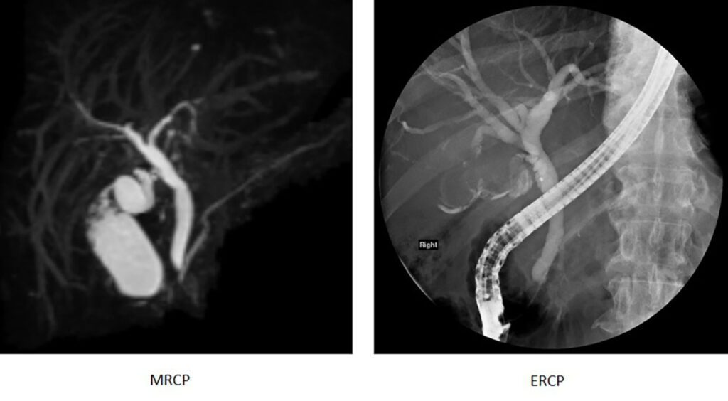
The fine-art of imaging the pancreato-biliary tree

A 50-year-old gentleman presents to his doctor with yellow skin, dark urine and pale stools. He also complains of poor appetite and unintentional weight loss of 10kg over 1 month. Blood tests reveal deranged liver function and raised tumour markers namely Ca 19-9 and CEA. MRI liver, pancreas and MR cholangiopancreatography (MRCP) was requested to investigate the cause. MRI showed a large growth in the liver originating from the bile duct, collectively measuring 12 cm – it is cancer. This was confirmed on tissue diagnosis. The patient underwent chemotherapy and passed away 8 months later.

The advent of MRCP

MRCP is a non-invasive technique used to image the bile ducts and pancreatic duct for evaluation of pancreatobiliary disease. It is a competitive substitute for endoscopic retrograde cholangiopancreatography (ERCP) especially in current times, with newer MR machines being able to produce good quality imaging through increased signal to noise ratio. Apart from evaluation of bile duct disease, MRCP is also able to evaluate for disease outside the bile ducts, for example, in the liver, when compared to ERCP. When MRCP is pitched against ultrasound, it triumphs as it is far more sensitive and less operator dependent.

Clinical Indications of MRCP

MRCP is indicated in the diagnosis of many benign (non-cancerous) and malignant (cancerous) conditions that affect the bile ducts and pancreas. It is commonly used look for the following conditions:-

Biliary tree

  • Check for anatomical variants of the biliary tree before surgery
  • Choledochal cyst
  • Bile duct stone
  • Benign (non-cancerous) bile duct narrowing from inflammation
  • Malignant (cancerous) bile duct narrowing from bile duct cancer (cholangiocarcinoma)

Gallbladder

  • Gallbladder disease such as gallstones

Pancreas

  • Annular pancreas
  • Pancreas divisum
  • Intraductal papillary mucinous neoplasms (IPMNs)
  • Pancreatic cancer

Technique

The high water content in bile composition and its relative stasis in the bile ducts are unique properties that are exploited to aid visualisation in MRCP imaging. Thin-slices of the bile ducts as well as multiple imaging planes are obtained over 30-40 minutes. These images will then be analysed and reported by a radiologist, alongside the patient’s clinical and surgical background.

To optimise an MRCP study, pineapple juice and black tea which contain manganese, have been used but not routinely, by nulling the bright signal in the GI tract that may obscure the biliary tree. For better visualisation of the pancreatic duct, synthetic secretin, a hormone, has been injected to produce temporary distension of the pancreatic ducts.

Most importantly, patients play a vital role in the MRCP examination since they are responsible for co-operation by holding their breath and/ or staying still during the study.

Conclusion

MRCP has evolved considerably over the last 2 decades with technological advances in both image acquisition and post-processing, resulting in significant improvement in its diagnostic capability. Whilst it will never replace the therapeutic capabilities of ERCP, it has been an extremely useful non-invasive examination in evaluation of pancreato-biliary disorders and adjacent organs such as the liver, like in our patient. In many cases it can be the only diagnostic study needed to provide an appropriate diagnosis.
Share this
Facebook
Twitter
LinkedIn
More Articles