
Background
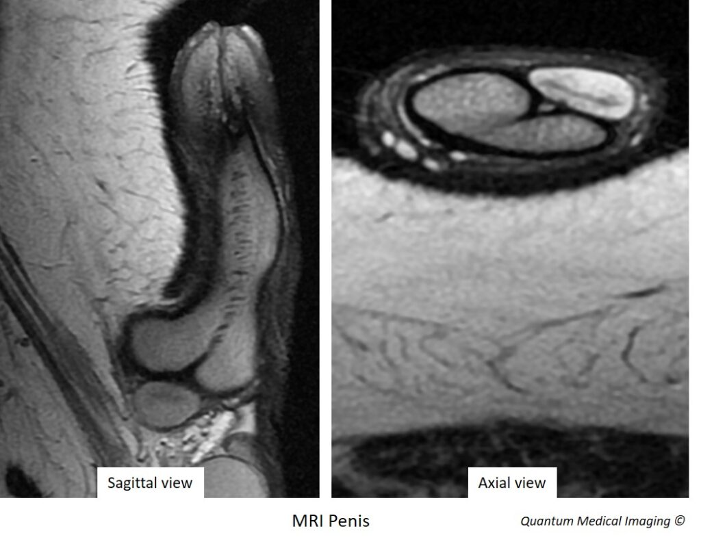
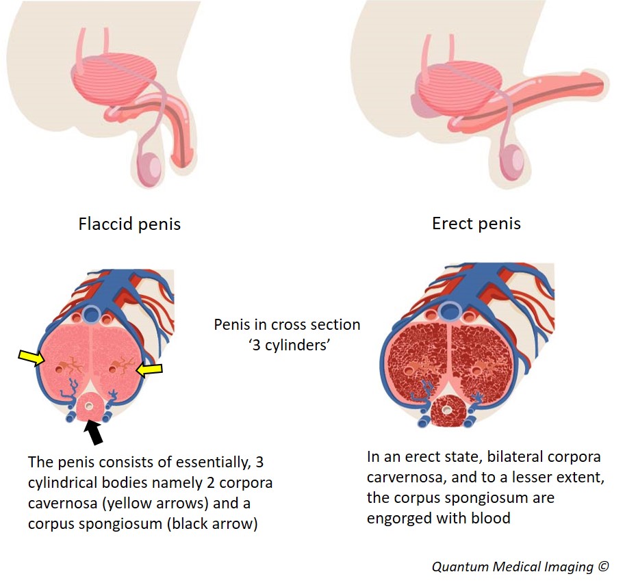
Anatomy of the penis
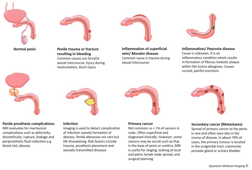
Clinical indications for penile MRI
- Trauma/ fracture of the penis
- Inflammation
- Infection
- Cancer
- Penile prosthesis complications
Technique
Appropriate positioning is essential for imaging the penis. The patient is placed lying on his back with feet first into the MRI scanner. The penis is usually taped to the front and midline against the front of the abdominal wall before it is scanned. The scan takes about 45 minutes.
The penis may be imaged in both flaccid and erect states. Imaging an erect penis improves visualisation of penile anatomy.
If imaging in an erect state is desired, injection of a prescribed synethetic hormone called prostaglandin E1 (Caverject) before the scan is performed. It takes approximately 10-20 minutes for the injection to work. There are instances where this injection cannot be given, that is, if the patient has a penile implant, acute penile fracture, painful erection and priapism. Alternative methods are to administer Sildenafil (Viagra) or manual stimulation.
Conclusion
MRI can contribute useful information for many different diseases of the male member but in many cases, not convincingly superior to clinical examination and US to justify its routine use. It is probably most useful in penile cancer staging, evaluation of penile prosthesis complications and imaging complex cases of fibrosis.
References
Lindquist CM et al. MRI of the penis. Abdominal Radiology (NY). 2020 Jul;45 (7):2001-2017. Springer. PubMed PMID: 31701192.
Kirkham A. MRI of the penis. Br J Radiol. 2012 Nov;85 Spec No 1(Spec Iss 1):S86-93. PubMED PMID: 23118102.
Long H. Tu, Spektor M, Ferrante M, Mathur M. MRI of the Penis: Indications, Anatomy, and Pathology. Review. Curr Probl Diagn Radiol. Jan-Feb 2020;49(1):54-63. PubMED PMID: 30704768


